
HALO Clinical AI Solutions


![]()
Indica Labs’ HALO is by far the most accurate, versatile, and user friendly digital image analysis platform that I have used. … HALO is a must have for any pathologist or scientist involved in quantifying immunohistochemical stains. In addition the HALO AI module is extremely easy to use. Within 20 minutes I was able to set up AI detection of tumor vs. normal tissue, as well as tumor epithelium vs stroma. HALO provides everything one needs for quantification of IHC for clinical and pre-clinical studies (of course with supervision of a pathologist). Best of all: it is fun, like a video game for Pathologists!
Senior Pathologist, AstraZeneca
![]()
Using HALO AP has made an enormous difference to the way I work. It is much easier to review complex cases and consult with colleagues. Frankly, if you took away my digital pathology system I’d probably retire, the reason being I can do things faster and more accurately than I can with glass.
Dr. David Clark, MD
Clinical Lead for Digital Pathology Implementation, Nottingham University Hospitals NHS Trust

The partnership with Indica Labs, a market leader in digital pathology, with interoperable workflow integration, will enable pathologists and labs to deliver faster, more accurate, and more reproducible answers using Lunit’s AI solution.
Dr. Brandon Suh, MD
CEO, Lunit

HALO is a user-friendly, and invaluable for quantification of multi-omics imaging data (MIBI, CODEX/Akoya multiplex stains, RNA-ISH/RNAscope). Cell segmentation, quantitation of tissue microarray data, aligning serial images, exporting quantitative data and .fcs files, it is incredible. The support team is incredibly responsive and collaborative.
Dr. Michael Kattah, MD, PhD
Assistant Professor, University of California, San Francisco


An Interview with Billy Heseltine, Director of Cloud Services – Indica Labs
An Interview with Billy Heseltine, Director of Cloud Services – Indica Labs

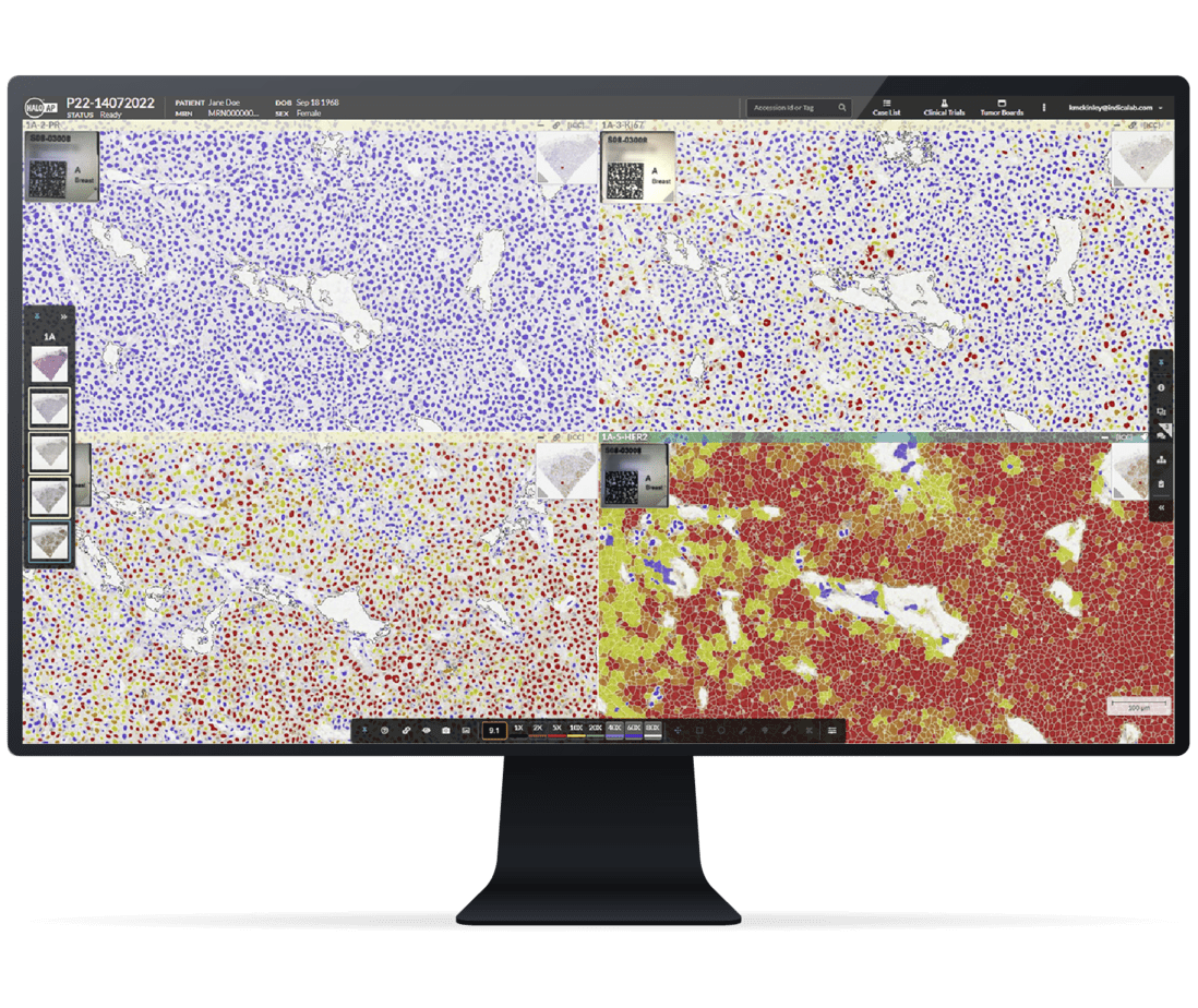
Integrating Innovation: HALO AP® as an AI Orchestration Platform for Digital Pathology
HALO AP seamlessly integrates with a wide selection of AI and image analysis innovations, LIS | LIMS, and diverse software platforms, offering unparalleled flexibility for your clinical workflow.

HALO Macrodissection Solutions: Enhancing the Accuracy of Personalized Oncology Through AI-Powered Slide Macrodissection
Lung Macrodissect AI identifies and highlights non-small cell lung cancer cells present on whole slide images to guide annotations for downstream automated slide macrodissection.

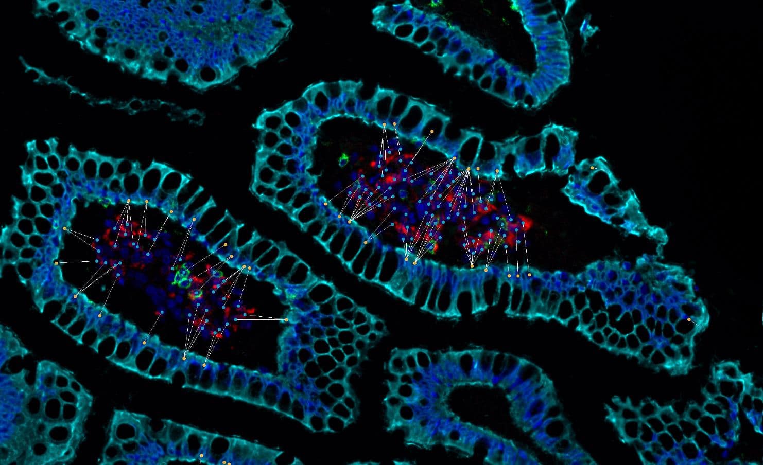
Announcing HALO® RNAscope™ modules supporting interactive markups
In this blog post, you can learn about the interactive markup image support added to the HALO® ISH, FISH, ISH-IHC, and FISH-IF modules, where to find the user guides, tutorial videos, and when to expect to upgrade.


Pathologist-Trained Machine Learning Classifiers Quantitate Celiac Disease Features
30 May 2024 | Histologic evaluation of the mucosal changes associated with celiac disease is important for establishing an accurate diagnosis and monitoring the impact of investigational therapies.

Indica Labs’ Boston HALO® User Group Meeting
16 May 2024 | Indica Labs is pleased to announce our Boston HALO® User Group Meeting at Le Méridien Boston Cambridge on May 16 from 1 pm – 5 pm. A lunch will be provided to all pre-registered attendees at 12 pm.

AI-Powered Pathology with HALO®, HALO AI, and HALO Link 4.0
6 June 2024 | Join us for this 1-hour webinar to see a live demonstration of the new versions of the industry leading AI-powered HALO® and HALO AI digital pathology image analysis platform and the HALO Link collaborative image management platform.
Masterclass Webinar: Intro to the HALO AI Toolbox
29 June 2023 | Join us for this 1-hour webinar to see a breakdown of the networks that make up HALO AI and applications suitable for each one.
Choosing The Right Foundation: Selecting And Deploying An Image Management System For Diagnostic Digital Pathology
18 May 2023 | In this 60-minute webinar, we will discuss considerations when selecting a digital pathology image management system for clinical diagnostics. In the first half of the event, we pose questions and suggest approaches that your group may adopt when evaluating diagnostic digital pathology platforms. A comprehensive Vendor
Transforming Pathology at Nottingham University Hospitals: Implementing Digital Pathology using HALO AP®
2 March 2023 | Learn about transformational change in how pathology services are delivered at Nottingham University Hospitals
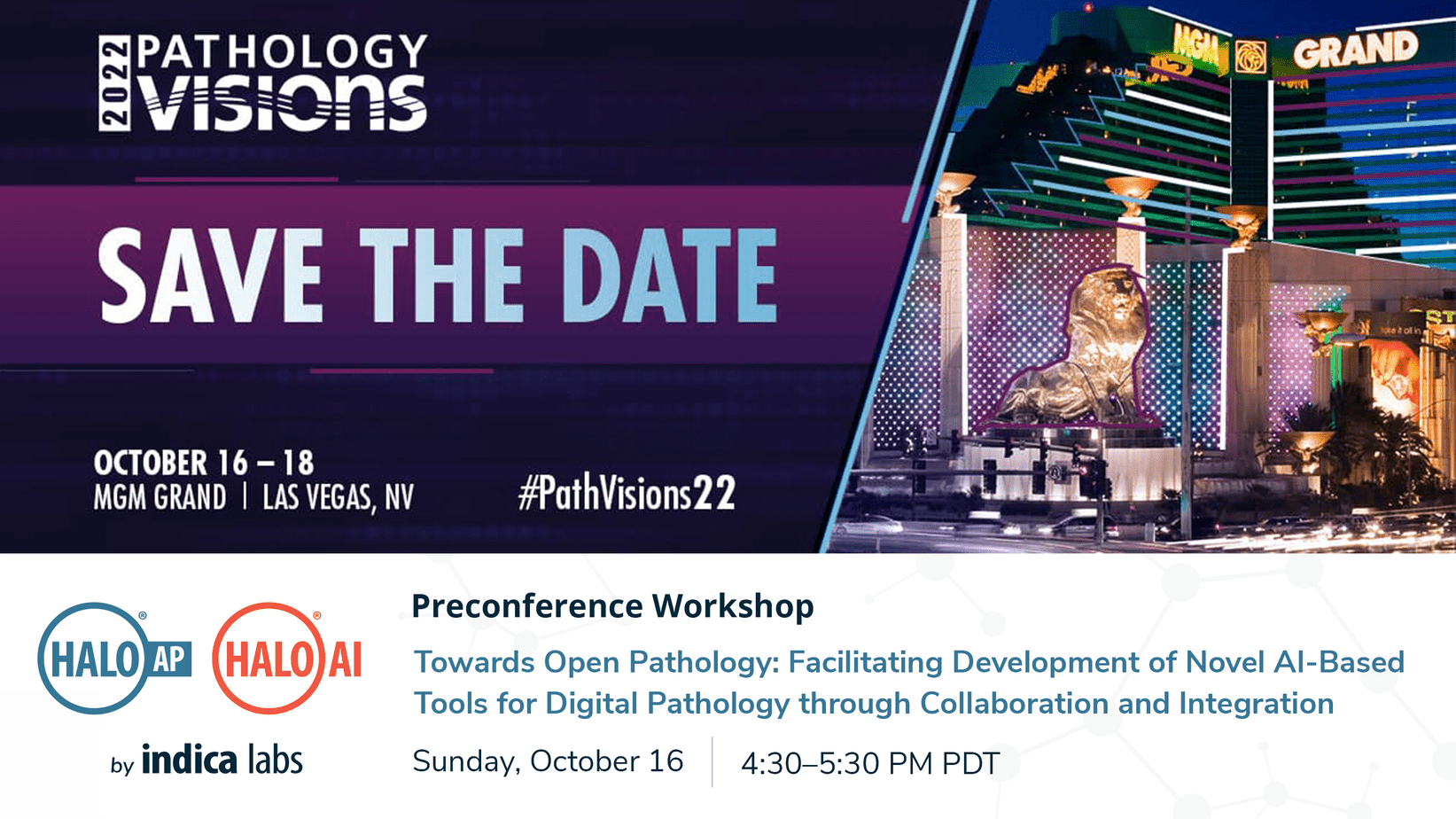
Towards Open Pathology: Facilitating Development of Novel AI-based Tools for Digital Pathology through Collaboration and Integration
16 October 2022 | In this 1-hour workshop, Eric Runde, Chief Operating Officer at Indica Labs, will introduce Indica’s platforms, our collaborative approach to incorporating artificial intelligence (AI) into life science and clinical workflows, and the open pathology tools available to developers and industry partners. Dr. Peter Caie, Principal Scientist
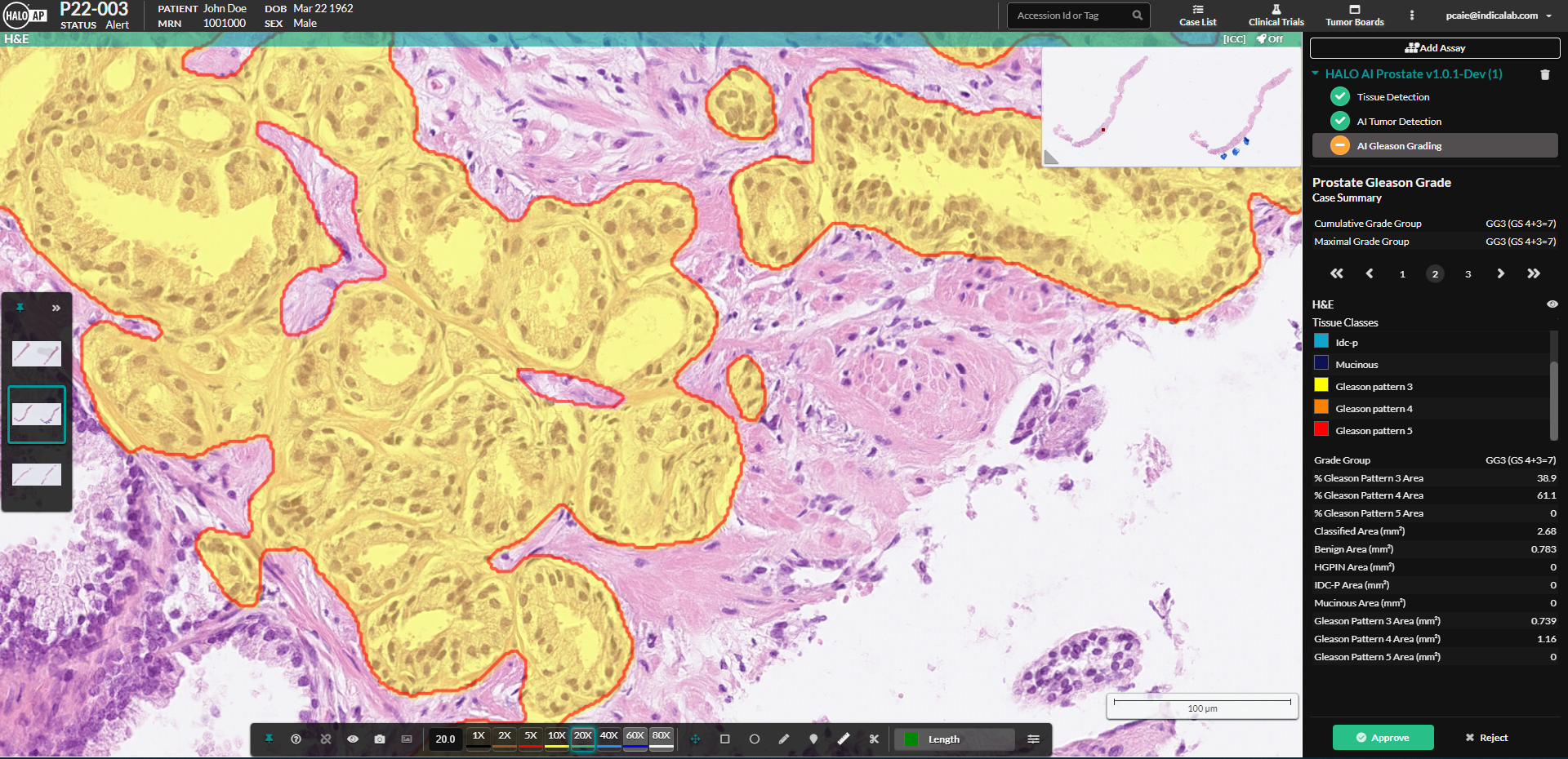
HALO Prostate AI – A Tool for Automated Detection and Gleason Grading of Prostate Cancer
05 October 2022 | Join us for this 1-hour webinar to learn how to implement the HALO Prostate AI tool for prostate cancer detection and grading
Quantitative Digital Pathology: An Overview of the HALO® Image Analysis Platform
6 October 2022 | Join us for this 1-hour webinar to see a live demonstration of the HALO® image analysis platform, the gold standard analysis platform for quantitative tissue analysis in digital pathology.
