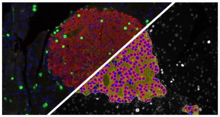
Islet FL
Count pancreatic islets fluorescently-labeled with up to three dyes, quantify number of dye positive cells per islet and area of dye positivity per islet.
Count pancreatic islets fluorescently-labeled with up to three dyes, quantify number of dye positive cells per islet and area of dye positivity per islet.
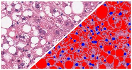
Quantifies vacuole area, diameter, perimeter, and number of vacuoles per region of interest. Measures steatosis in kidney, lipid droplets in adipose tissue, and alveolar area in lung tissue.
Vacuole Quantification Read More »
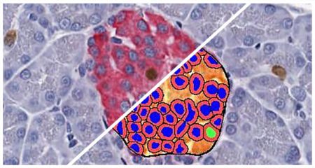
Count pancreatic islets, quantify number of alpha and beta cells in each islet, and determine area of islets stained with insulin and glucagon or other islet-specific stains
Islet Quantification Read More »
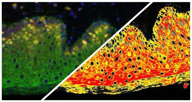
Measure the positive area, average intensity and dye colocalization (where applicable) of an unlimited number of fluorescent dyes.
Area Quantification FL Read More »

Deconvolve up to five colors in brightfield and measure positive area and average optical density for each stain and stain colocalization (where applicable).
Area Quantification Read More »
Courtney M. Williams et al, The FASEB Journal, 2021
Williams and colleagues at Regeneron Pharmaceuticals perform structure/function relationship studies on Fgf15 and FGF19 using site-directed mutagenesis and downstream functional assays in order to understand their distinct functions in a common pathway. Both molecules are therapeutic targets due to involvement in hepatocellular carcinoma and bile acid production. This publication identifies a single cysteine residue is identified that controls dimerization and hepatocyte proliferation. Understanding these molecular pathways may inform future studies on hepatocellular carcinoma while limiting toxicity and induction of hepatocyte proliferation. Immunohistochemistry images were quantified in HALO Link using the Cytonuclear IHC module.