


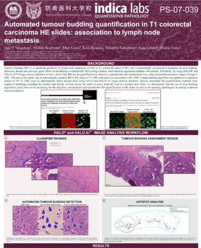
Quantify expression of up to five brightfield stains in any cellular compartment - membrane, nucleus or cytoplasm.
Learn More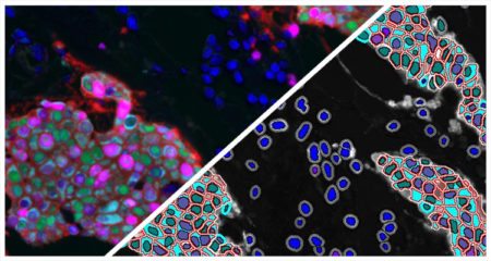
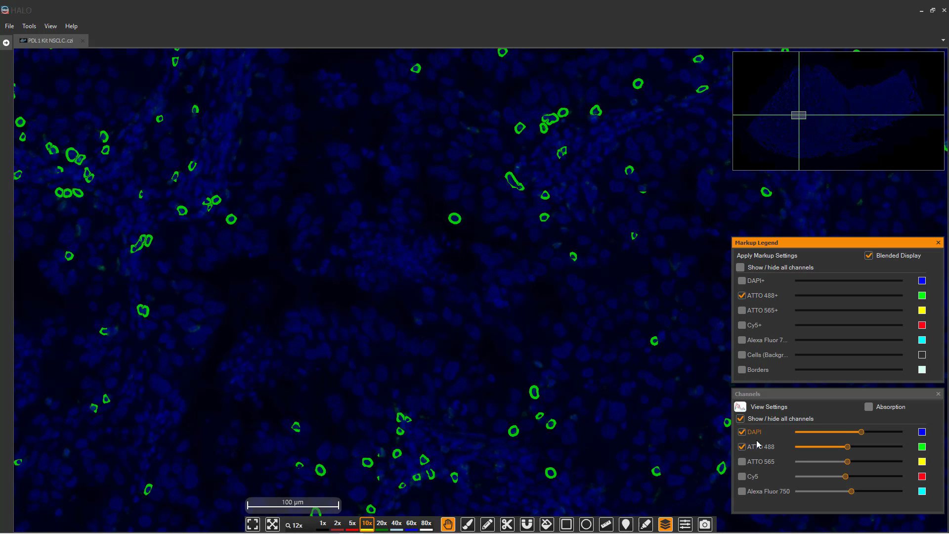
Quantify expression of an unlimited number of biomarkers in any cellular compartment - membrane, nucleus or cytoplasm.
Learn More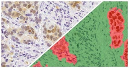
Separate multiple tissue classes across a tissue using a learn-by-example approach. Can be used in conjunction with all other modules (fluorescent and brightfield) to select specific tissue classes for further analysis.
Learn More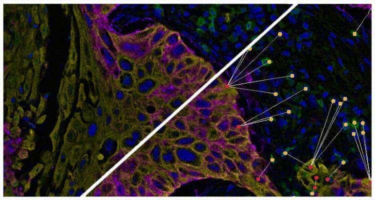
Plot cells and objects from one or more images and perform nearest neighbor analysis, proximity analysis, and tumor infiltration analysis.
Learn More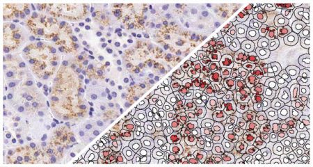
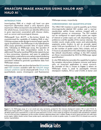
Quantify spot number, optical density and co-expression of one or two brightfield RNA or DNA probes on a per cell basis. Includes support for brightfield RNAscope assays.
Learn More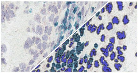
Quantify IHC positivity and spot number, optical density and co-expression of one or two brightfield RNA or DNA probes on a per cell basis. Includes support for brightfield RNAscope assays.
Learn More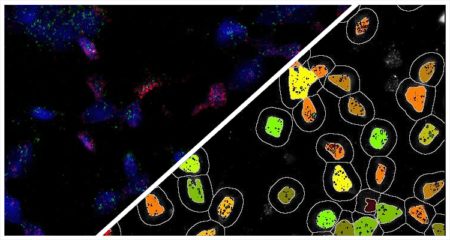
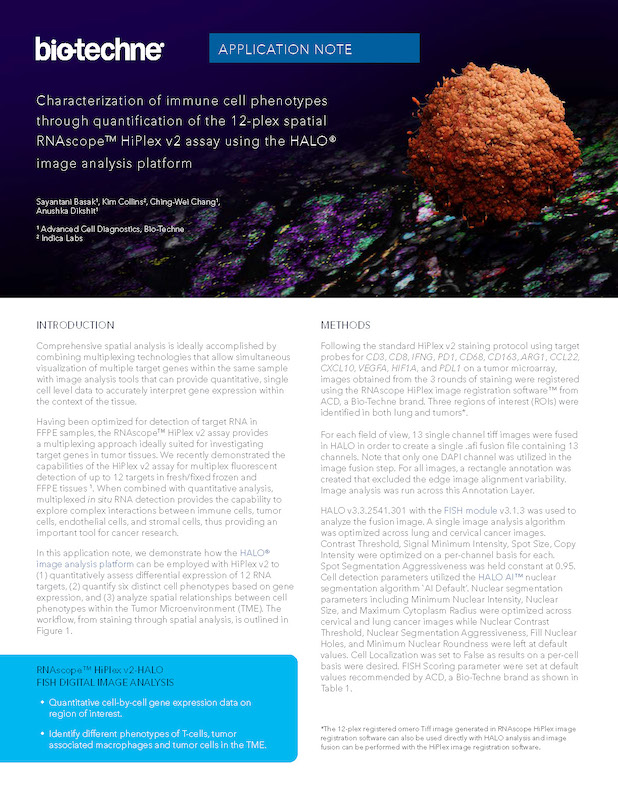
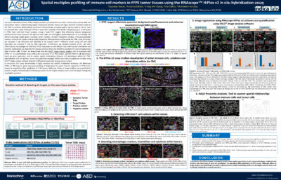
Quantify number, intensity and co-expression of an unlimited number of fluorescently labeled RNA or DNA probes on a per cell basis. Includes support for fluorescent RNAscope assays.
Learn More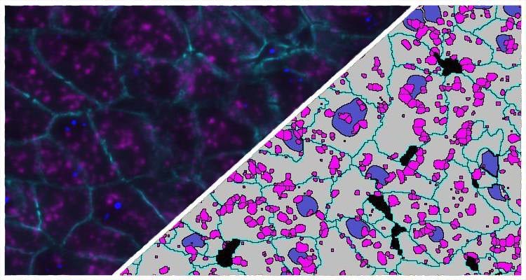
Quantify an unlimited number of fluorescent RNA/DNA probes and IF protein biomarkers on a per cell basis. Includes support for fluorescent RNAscope assays.
Learn More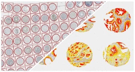
Module to assist in the segmentation of TMA spots for further analysis.
Learn More


SARS-CoV-2, the causative agent of COVID-19, has been the focus of intense research since its detection in late 2019. It is known that viral infection affects several cellular processes, including senescence and inflammation, and prior studies also point toward SARS-CoV-2...
Learn More
Schwannomas are sporadic, usually benign tumors that develop predominately on spinal nerve roots and cranial nerves. Inactivation of NF2 in Schwann cells (SC) is the cause of nearly all schwannomas and mutations in additional genes are few if present at...
Learn More
Stereotactic body radiotherapy (SBRT) uses multiple targeted beams of radiation to damage malignant cells while limiting effects to surrounding tissue. SBRT is a standard treatment for various cancers that elicit strong immune responses, but immunologically cold tumors, such as pancreatic...
Learn More

Pathologist-Trained Machine Learning Classifiers Quantitate Celiac Disease Features
30 May 2024 | Histologic evaluation of the mucosal changes associated with celiac disease is important for establishing an accurate diagnosis and monitoring the impact

AI-Powered Pathology with HALO®, HALO AI, and HALO Link 4.0
6 June 2024 | Join us for this 1-hour webinar to see a live demonstration of the new versions of the industry leading AI-powered HALO®
HALO AI 4.0 Sneak Peek
29 February 2024 | Please join us for this 1-hour webinar to learn about the exciting new features coming soon in HALO AI 4.0!

HALO® 4.0 Sneak Peek
15 February 2024 | Please join us for this 1-hour webinar to learn about the exciting new features coming soon in HALO 4.0!

Indica Labs and Molecular Instruments Announce Partnership to Advance AI-Powered Quantitative Image Analysis with HCR™ RNA-ISH
Albuquerque, NM, and Los Angeles, CA – 5 April 2024 – Indica Labs, a leading provider of AI-powered digital pathology solutions, and Molecular Instruments® (MI),

Indica Labs Launches Comprehensive Management Service for HALO® Software in AWS Cloud-Hosted Environments
Albuquerque, NM – 8 June 2023 – Indica Labs today announces the launch of a service offering deployment and on-going support of its HALO® digital pathology
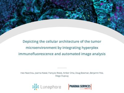
Press Release: Lunaphore and Indica Labs announce partnership to provide complete technology solution for spatial biology and image analysis
LAUSANNE, Switzerland and ALBUQUERQUE, N.M. – May 12, 2022 – 5 pm (CET) | 9 am MDT – Lunaphore, a Swiss life sciences company developing
Press Release: Indica Labs announces launch of enterprise-wide, cloud-based digital pathology deployment at NCI
Albuquerque, NM – October 8, 2020 – Indica Labs, a leading provider of computational pathology software and services, are pleased to announce the formal launch

Announcing HALO® RNAscope™ modules supporting interactive markups
In this blog post, you can learn about the interactive markup image support added to the HALO® ISH, FISH, ISH-IHC, and FISH-IF modules, where to
Looking Back: Reviewing 2023 at Indica Labs
As we welcome the new year and look back on 2023, the Indica Labs team wants to extend our heartfelt thanks to our customers and
Taking HALO® to the Cloud with Indica Labs Cloud Services
In this blog post we will discuss the basics of cloud computing, benefits and considerations of cloud vs. traditional deployments, and how our Professional
Indica Labs Launches Comprehensive Management Service for HALO® Software in AWS Cloud-Hosted Environments
Albuquerque, NM – 8 June 2023 – Indica Labs today announces the launch of a service offering deployment and on-going support of its HALO® digital pathology

Indica Labs’ Boston HALO® User Group Meeting
16 May 2024 | Indica Labs is pleased to announce our Boston HALO® User Group Meeting at Le Méridien Boston Cambridge on May 16 from

Indica Labs’ Frankfurt HALO® User Group Meeting
Indica Labs is pleased to announce our Frankfurt HALO® User Group Meeting at Hyatt Place Frankfurt Airport Hotel on 13 March from 12:00 – 18:00.

Indica Labs’ London HALO® User Group Meeting
6 December 2023 | Indica Labs is pleased to announce our London HALO® User Group Meeting to be held in London on 6 December 2023

Japan User Group Meeting
Indica Labs’ Japan HALO® User Group Meeting Date: 20 October 2023 Time: 10:30 – 16:00 (GMT+8) Location: Senri Room A, Senri Life Science Center, Osaka,
