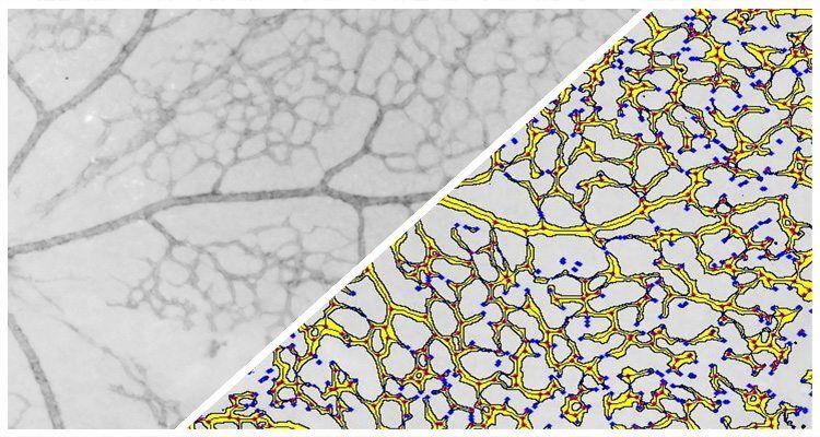
Branch Structure FL
Analyze branch-like structures such as retinal vessels or cortical neurons in fluorescence.
Branch Structure FL Read More »
Analyze branch-like structures such as retinal vessels or cortical neurons in fluorescence.
Branch Structure FL Read More »
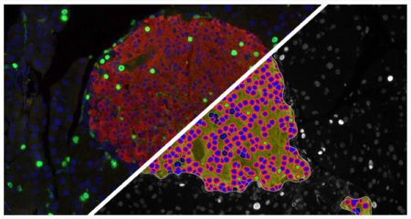
Count pancreatic islets fluorescently-labeled with up to three dyes, quantify number of dye positive cells per islet and area of dye positivity per islet.
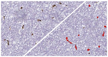
Count one or two objects and measure the object area, diameter, stain intensity, and colocalization (where applicable). Microvessel and amyloid plaque quantification are most common applications.
Object Colocalization Read More »
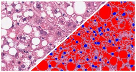
Quantifies vacuole area, diameter, perimeter, and number of vacuoles per region of interest. Measures steatosis in kidney, lipid droplets in adipose tissue, and alveolar area in lung tissue.
Vacuole Quantification Read More »
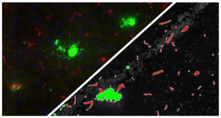
Count one or two fluorescently labeled objects and measures the object area, diameter, stain intensity, and colocalization (where applicable). Microvessel and amyloid plaque quantification are most common applications.
Object Colocalization FL Read More »
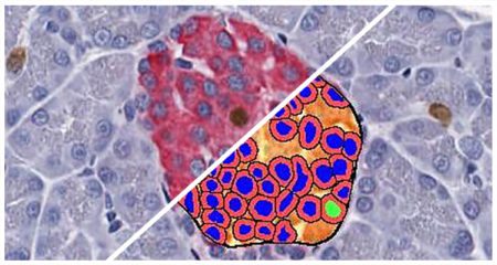
Count pancreatic islets, quantify number of alpha and beta cells in each islet, and determine area of islets stained with insulin and glucagon or other islet-specific stains
Islet Quantification Read More »
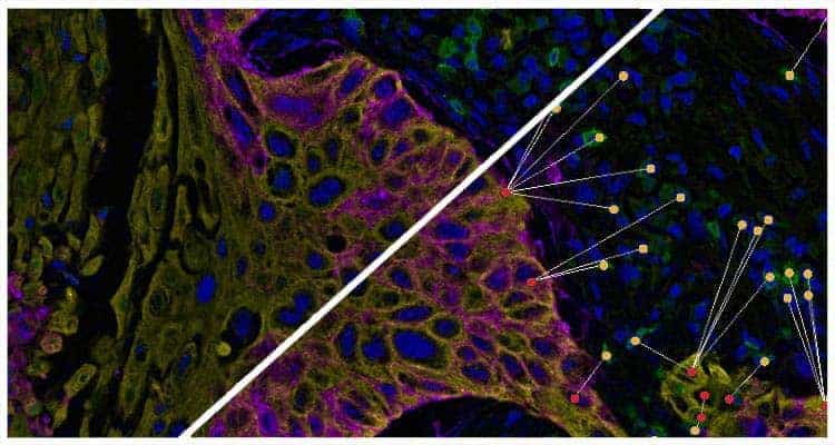
Plot cells and objects from one or more images and perform nearest neighbor analysis, proximity analysis, and tumor infiltration analysis.
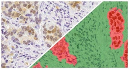
Separate multiple tissue classes across a tissue using a learn-by-example approach. Can be used in conjunction with all other modules (fluorescent and brightfield) to select specific tissue classes for further analysis.
Tissue Classifier Add-On Read More »